NCERT Solutions For Class 11 Biology Neural Control and Coordination
Topics and Subtopics in NCERT Solutions for Class 11 Biology Chapter 21 Neural Control and Coordination:
| Section Name | Topic Name |
| 21 | Neural Control and Coordination |
| 21.1 | Neural System |
| 21.2 | Human Neural System |
| 21.3 | Neuron as Structural and Functional Unit of Neural System |
| 21.4 | Central Neural System |
| 21.5 | Reflex Action and Reflex Arc |
| 21.6 | Sensory Reception and Processing |
| 21.7 | Summary |
NCERT Solutions Class 11 BiologyBiology Sample Papers
NCERT TEXTBOOK QUESTIONS FROM SOLVED
1. Briefly describe the structure of the following:
(a) Brain (b) Eye (c) Ear
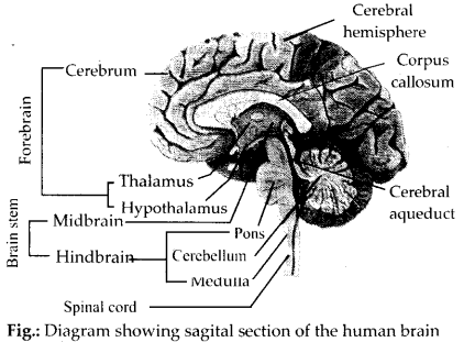
Solution: (a) Brain: The brain acts as control and command system of the body. It is protected by skull and is covered by three meninges. It is divisible into three main regions: forebrain, midbrain and hindbrain.
(i) Forebrain – It consists of three regions:
(a) Olfactory lobes: These are a pair of very small, solid club-shaped bodies which are widely separated from each
other. They are fully covered by cerebral hemispheres.
(b) Cerebrum – It is the largest and most complex of all the parts of human brain. A deep cleft divides the cerebrum into right and left cerebral hemispheres, connected by myelinated fibres, the corpus callosum.
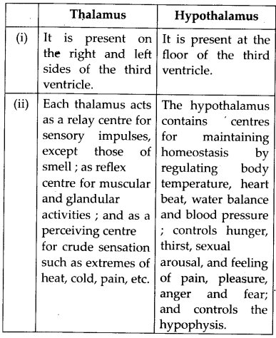
(c) Diencephalon – It encloses a slit-like cavity, the third ventricle. The thin roof of this cavity is known as the epithalamus, the thick right and left sides as the thalami, and floor as the hypothalamus.
(ii) Midbrain – It is located between thalamus/ hypothalamus of forebrain and pons of hindbrain. Its upper surface has two pairs of rounded protrusious called corpora quadrigemina and two bundles of fibres called crura cerebri.
(iii) Hindbrain – It consists of:
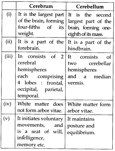
(a) Cerebellum – The second largest part of the human brain is the cerebellum. It consists of two lateral cerebellar hemispheres and central worm-shaped part, the vermis. The cerebellum has its grey matter on the outside, comprising three layers of cells and fibres. It also has Golgi cells, basket cells and granule cells.
(b) Pons varolii – An oval mass, called the pons varolii, lies above the medulla oblongata. It consists mainly of nerve fibres which interconnect different regions of the brain.
(c) Medulla oblongata – It extends from the pons varolii above and is continuous with the spinal cord below. The mid brain, pons varolii and medulla oblongata are collectively called brain stem.
(b) Eye: Eye is a hollow spherical structure composed of three coats:
– Outer fibrous coat
– Middle vascular coat
– Inner nervous coat
(i) Fibrous coat: It is thick and protects the eyeball. It has two distinct regions – sclera and cornea. Sclera covers most of the eye ball. The sclera or white of the eye contains many collagen fibres. Cornea is a transparent portion that forms the anterior one – sixth of the eyeball. The cornea is avascular (i.e., lacks blood supply).
(ii)Vascular coat: It comprises of 3 regions : choroid, iris, ciliary body.
(a) Choroid : It lies adjacent to sclera and contains numerous blood vessels and pigmented cells.
(b) Iris: The iris is a circular muscular diaphragm containing the pigment giving eye its colour. It extends from the ciliary body across the eyeball in front of the lens. It 2. has an opening in the centre called the pupil.
It contains two types of smooth muscles, circular muscles (sphincters) and radial muscles (dilators), of ectodermal origin.
(c) Ciliary body: Behind the peripheral margin of the iris, the vascular coat is thickened to form the ciliary body. It is composed of the ciliary muscles and the ciliary processes.
(iii) Nervous coat: It consists of retina which is neural and sensory layer of an eye ball. It consists of three layers; ganglion cells, bipolar cells and photoreceptor cells (rods and cones).
Lens: It is a transparent, biconvex, elastic structure that bends light waves as they pass through its surface. It is composed of epithelial cells that have large amounts of clear cytoplasm in the form of fibres.
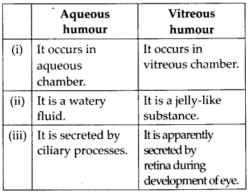
Chambers of eyeball: The lens, suspensory ligament and ciliary body divide the eye into an anterior aqueous chamber and a posterior vitreous chamber which are filled with aqueous humour and vitreous humour respectively.
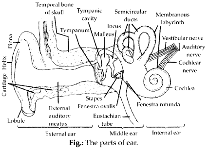
(c) Ear: There are three portions in an ear:
(i) External ear: It further has 2 regions: pinna and external auditory canal or meatus.
(a) Pinna: The pinna is a projecting elastic cartilage covered with skin. Its most prominent outer ridge is called the helix. The lobule is the soft pliable part at its lower end composed of fibrous and adipose tissue richly supplied with blood capillaries. It is sensitive as well as effective in collecting sound waves.
(b) External auditory canal: It is an S-shaped tube leading inward from the pinna. It is a tubular passage supported by cartilage in its exterior part and by bone in its interior part.
(ii) Middle ear: It consists of 3 small bones called ear ossicles – malleus, incus and stapes, which are attached to one another and increase efficiency of transmission of sound waves to inner ear.
(iii) Internal ear: It consists of bony and
2. Compare the following:
(a) Central neural system (CNS) and Peripheral neural system (PNS).
(b) Resting potential and action potential.
(c) Choroid and retina.
Solution: (a) CNS: It lies along the mid-dorsal axis of the body. It is a hollow, dorsally placed structure and comprises of brain and spinal cord. It is a centre of information processing and control.
PNS: Nerves arising from the central nervous system constitute the peripheral nervous system. It carries information to and from the CNS. It includes spinal nerves and cranial nerves.
(b) Resting potential: Outside the plasma membrane of a nerve fibre is the extracellular fluid which is positively charged with respect to the cell contents inside the plasma membrane. A resting nerve fibre shows a potential difference between inside and outside of this plasma membrane. This difference in the electrical charges across the plasma membrane is called the ‘resting potential’. A membrane with resting potential across it, is said to be electrically polarized. Action potential : Action potential is another name of nerve impulse. The contents inside a cell at the excited state becomes positively charged with respect to extracellular fluid outside it. This change in polarity across the plasma membrane is known as an action potential. The membrane with reversed polarity across it is said to be depolarized.
(c) Choroid: Choroid lies adjacent to the sclera and contains numerous blood vessels that supply nutrients and oxygen to the other tissues especially of retina. It contains abundant pigment cells and is dark brown in colour.
Retina: It is the neural and sensory layer of the eye ball. It is a very delicate coat and lines the whole of the vascular coat. Its external surface is in contact with the choroid and its internal surface with vitreous humour. It contains ganglion cells, bipolar cells and photoreceptor cells. membranous labyrinth. Membranous labyrinth consists of three semicircular ducts, utricle, saccule and cochlea.
More Resources for CBSE Class 11
- NCERT Solutions
- NCERT Solutions Class 11 Maths
- NCERT Solutions Class 11 Physics
- NCERT Solutions Class 11 Chemistry
- NCERT Solutions Class 11 Biology
- NCERT Solutions Class 11 Hindi
- NCERT Solutions Class 11 English
- NCERT Solutions Class 11 Business Studies
- NCERT Solutions Class 11 Accountancy
- NCERT Solutions Class 11 Psychology
- NCERT Solutions Class 11 Entrepreneurship
- NCERT Solutions Class 11 Indian Economic Development
- NCERT Solutions Class 11 Computer Science
3. Explain the following processes:
(a) Polarisation of the membrane of a nerve fibre.
(b) Depolarisation of the membrane of a nerve fibre.
(c) Conduction of a nerve impulse along a nerve fibre.
(d) Transmission of a nerve impulse across a chemical synapse.
Solution: (a) Polarisation of the membrane of a nerve fibre : In the resting (not conducting impulse) nerve fibre the plasma membrane separates two solution of different chemical composition but having approximately the same total number of ions. In the external medium (tissue fluid), sodium ions (Na+) and Cl– ions predominate, whereas within the fibre (intracellular fluid) potassium ions (K+) predominate. The differential flow of the positively charged ions and the inability of the negatively charged organic (protein) ions within the nerve fibre to pass out cause an increasing positive charge on the outside of the membrane and negative charge on the inside of the membrane. This makes the membrane of the resting nerve fibre polarized, extracellular fluid outside being electropositive (positively charged) with respect to the cell contents inside it.
(b) Depolarisation of the membrane of a nerve fibre: During depolarisation, the activation gates of Na channels open, and the K channels remain closed. Na+ rush into the axon. Entry of sodium ions leads to depolarisation (reversal of polarity) of the nerve membrane, so that the nerve fibre contents become electropositive with respect to the extracellular fluid.
(c) Conduction of a nerve impulse along a nerve fibre: Nervous system transmits information as a series of nerve impulses. A nerve impulse is the movement of an action potential as a wave through a nerve fibre. Action potentials are propagated, that is, self-generated along the axon. The events that set up an action potential at one spot on the nerve fibre also transmit it along the entire length of the nerve fibre. The action potential then moves to the neighbouring region of the nerve fibre till it covers the whole length of the fibre.
(d) Transmission of a nerve impulse across a chemical synapse: At a chemical synapse, the membranes of the pre- and post- synaptic neurons are separated by a fluid- filled space called synaptic cleft. Chemicals called neurotransmitters are involved in the transmission of impulses at these synapses. The axon terminals contain vesicles filled with these neurotransmitters. When an impulse (action potential) arrives at the axon terminal, it stimulates the movement of the synaptic vesicles towards the membrane where they fuse with the plasma membrane and burst to release their neurotransmitters in the synaptic cleft. The released neurotransmitters bind to their specific receptors, present on the post- synaptic membrane. This binding opens ion channels allowing the entry of ions which can generate a new potential in the post-synaptic neuron. The new potential developed may be either excitatory or inhibitory.
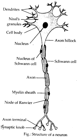
4. Draw labelled diagrams of the following:
(a) Neuron (b) Brain
(c) Eye (d) Ear
Solution: (a)
(b)
(c)

(d)
5. Write short notes on the following:
(a) Neural coordination (b) Forebrain
(c) Midbrain (d) Hindbrain
(e) Retina (f) Ear ossicles
(g) Cochlea (h) Organ of Corti
(i) Synapse
Solution: (a) Neural coordination : When higher animals respond to various stimuli, each response to a specific stimulus generally involves many organs (parts) of their bodies. Therefore, it is necessary that all the concerned organs (parts) of the body should work in a systematic manner to produce the response. The working together of various organs (parts) of the body of multicelullar organism in a proper manner to complement the functions of each other is called coordination. This is achieved by three overlapping processes of nervous system-sensory input, integration and motor output.
(b) Forebrain: It consists of: Olfactory lobes, the paired structures concerned with the sense of smell. Cerebrum which is the largest and most complex of all the parts of the human brain. It is divided by a cleft into left and right cerebral hemispheres which are connected by a large bundle of myelinated fibres the. corpus callosum. The outer cover of cerebral hemisphere is called cerebral cortex. It consists of sensory and motor areas. Hypothalamus region of forebrain contains centres which control body temperature, hunger and also contains group of neurosecretory cells.
(c) Midbrain: The midbrain is located between the thalamus/hypothalamus of the forebrain and pons of the hindbrain. A canal called the cerebral aqueduct passess through the midbrain. The dorsal portion of the midbrain consists mainly of four round swellings (lobes) called corpora quadrigemina. Midbrain and hindbrain form the brain stem.
(d) Hindbrain: The hindbrain comprises pons, cerebellum and medulla. Pons consists of fibre tracts that interconnect different regions of the brain. Cerebellum has very convoluted surface in order to provide the additional space of many more neurons. The medulla of the brain is connected to the spinal cord. The medulla contains centres which control respiration, cardiovascular reflexes and gastric secretions.
(e) Retina: Retina is the inner layer of an eye and it contains three layers of cells-from inside to outside – ganglion cells, bipolar cells and photoreceptor cells. There are two types of photoreceptor cells, namely, rods and cones. These cells contain the light-sensitive proteins called the photopigments. The daylight (photopic) vision and colour vision are functions of cones and the twilight (scotopic) vision is the function of the rods. The rods contain a purplish-red protein called the rhodopsin or visual purple, which contains a derivative of Vitamin A. In the human eye, there are three types of cones which possess their own characteristic photopigments that respond to red, green and blue lights. The sensations of different colours are produced by various combinations of these cones and their photopigments. When these cones are stimulated equally, a sensation of white light is produced.
(f) Ear ossicles : There is a small flexible chain of three small bones called as ear ossicles – the malleus (hammer shaped), the incus (anvil shaped) and the stapes (stirrup shaped) in the middle ear. Malleus is attached to the tympanic membrane on one side and incus on the other side. Incus in turn is connected with the stapes. Malleus is the largest ossicle, however stapes is the smallest ossicle.
(g) Cochlea : It is the main hearing organ which is connected with saccule. It is a spirally coiled tube that resembles a snail shell in appearance. It tapers from a broad base to an almost pointed apex.
(h) Organ of Corti: It is a structure located on the basilar membrane which contains hair cells that act as auditory receptors. The hair cells are present in rows on the internal side of the organ of Corti.
(i) Synapse : It is the junction between the axon of one neuron and the dendrite or cyton of another neuron for transmission of nerve impulse.
6. Give a brief account of
(a) Mechanism of synaptic transmission.
(b) Mechanism of vision.
(c) Mechanism of hearing.
Solution: (a) Mechanism of synaptic transmission: Refer answer 3 (d)
(a) Mechanism of vision: The light rays in visible wavelength focused on the retina through the cornea and lens generate potentials (impulses) in rods and cones. Light induces
dissociation of the retinal from opsin resulting in changes in the structure of the opsin. This causes membrane permeability changes. As a result, potential differences are generated in the photoreceptor cells. This produces a signal that generates action potentials in the ganglion cells through the bipolar cells. These action potentials (impulses) are transmitted by the optic nerves to the visual cortex area of the brain, where the neural impulses are analysed and the image formed on the retina is recognised based on earlier memory and experience.
(b) Mechanism of hearing : The external ear receives sound waves and directs them to the ear drum. The ear drum vibrates in response to the sound waves and these vibrations are transmitted through the ear ossicles (malleus, incus and stapes) to the oval window. The vibrations are passed through the oval window on to the fluid of the cochlea, where they generate waves in the lymphs. The waves in the lymphs induce a ripple in the basilar membrane. These movements of the basilar membrane bend the hair cells, pressing them against the tectorial membrane. As a result, nerve impulses are generated in the associated afferent neurons. These impulses are transmitted by the afferent fibres via auditory nerves to the auditory cortex of the brain, where the impulses are analysed and the sound is recognised.
7. Answer briefly.
(a) How do you perceive the colour of an object?
(b) Which part of our body helps us in maintaining the body balance?
(c) How does the eye regulate the amount of light that falls on the retina?
Solution: (a)In humans, colour vision results from the activity of cone cells, a type of photoreceptor cells. In the human eye, there are three types of cones which possess their own characteristic photopigments that respond to red, green and blue lights. The sensations of different colours are produced by various combinations of these cones and their photopigments. When these cones are stimulated equally, sensation of white light is produced. Yellow light, for instance, stimulates green’and red cones approximately to equal extent, and this is interpreted by the brain as yellow colour.
(b) Ears (cristae and maculae present in internal ears).
(c) The iris contains two sets of smooth muscles – sphincters and dilators. These muscles regulate the amount of light entering the eyeball by varying the size of pupil. Contraction of sphincter muscles makes the pupil smaller in bright light so that less light enters the eye. Contraction of dilator muscles widens the pupil in dim light so that more light goes in eye to fall on retina.
8. Explain the following.
(a) Role of Na+ in the generation of action potential.
(b) Mechanism of generation of light-induced impulse in the retina.
(c) Mechanism through which a sound produces a nerve impulse in the inner ear.
Solution: (a) The action potential is largely determined by Na+ ions. The action potential results from the following sequential events
(i) Disturbance caused to the membrane of a nerve fibre by a stimulus results in leakage of Na+ into the nerve fibre.
(ii) Entry of Na+ lowers the trans-membrane potential difference.
(iii) Decrease in potential difference makes the membrane more permeable to Na+ than to K+ ions so that more Na+ enter the fibre than K+ leave it.
(iv) Accumulation of Na+ in the nerve fibre initiates depolarisation (action potential), making the axonic contents positively charged relative to the extracellular fluid.
(v) With continued addition of Na+ the potential reaches zero and then plus 40-50 millivolts. This is the peak of action potential.
(vi) Permeability of a depolarised membrane to Na+ then rapidly drops, there are now as many Na+ on the inside of the membrane as on the outside.
(b) Refer answer 6 (b)
(c) Refer answer 6 (c)
9. Differentiate between
(a) Myelinated and non-myelinated axons
(b) Dendrites and axons
(c) Rods and cones
(d) Thalamus and Hypothalamus
(e) Cerebrum and Cerebellum
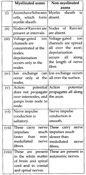
Solution: (a) Differences between myelinated and non-myelinated axons are as follows:
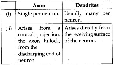
(b) Axon and dendrites can be differentiated as follows:
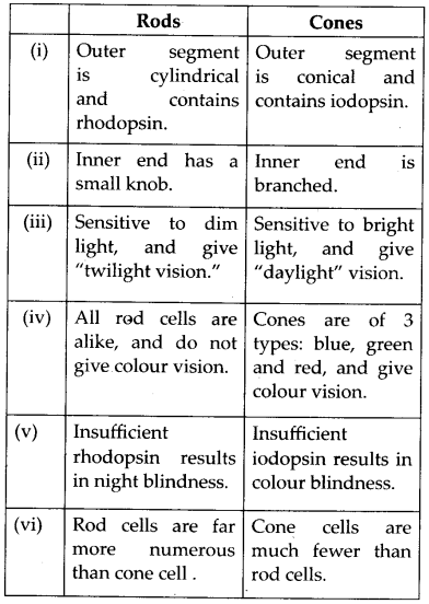
(c) The differences between rods and cones are as follows:
(d) Thalamus and hypothalamus can be differentiated as follows:
(e) Cerebrum and cerebellum can be differentiated as follows:
10. Answer the following.
(a) Which part of the ear determines the pitch ofa sound?
(b) Which part of the human brain is the most developed?
(c) Which part of our central neural system acts as a master clock?
Solution: (a) The receptor cells in the organ of Corti (Internal ear).
(b) Cerebrum (cerebral hemispheres).
(c) Pineal gland present in diencephalon of forebrain acts as a master clock, which maintains biological rhythm.
11. The region of the vertebrate eye, where the optic nerve passes out of the retina, is called the
(a) fovea (b) iris
(c) blind spot (d) optic chiasma
Solution: (c) blind spot
12. Distinguish between
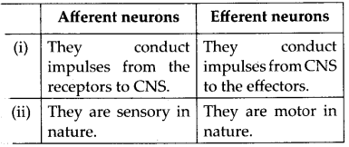
(a) Afferent neurons and efferent neurons
(b) Impulse conduction in myelinated nerve fibre and unmyelinated nerve fibre
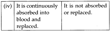
(c) Aqueous humour and vitreous humour
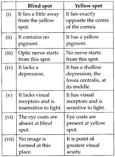
(d) Blind spot and yellow spot
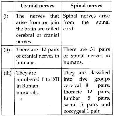
(e) Cranial nerves and spinal nerves
Solution: (a)
(b) Refer answer 9(a)
(c)
(d)
(e)